
Hyodeoxycholic acid reduces excess liver fat in mice through two distinct pathways, demonstrating multi-target therapeutic potential
Sept 17: Excess fat accumulation in the liver can contribute to the development and progression of metabolic dysfunction-associated steatotic liver disease (MASLD), highlighting the need for new approaches to manage the condition. Now, researchers have found that hyodeoxycholic acid (HDCA), a bile acid, can reduce liver fat in mice while also identifying the biological pathways involved. The findings suggest that HDCA could help inform the development of new approaches to managing MASLD.
Non-alcoholic fatty liver disease (NAFLD), now known as metabolic dysfunction-associated steatotic liver disease (MASLD), is a common condition that causes excess fat to build up in the liver, affecting about one-quarter of adults worldwide. In some people, this can lead to inflammation and liver cell or hepatocyte injury, triggering the formation of thick, fibrous connective tissue known as fibrosis. Over time, advanced fibrosis can progress to cirrhosis, in which permanent scarring impairs the liver’s normal function. MASLD is often described as a “silent” disease because symptoms may be absent or mild until the disease becomes more advanced.
Hepatic steatosis refers to the initial stage of MASLD, where excess fat gets accumulated in the hepatocytes. Though it is a common metabolic disorder, the mechanistic understanding of the biological pathways involved as well as effective therapeutic strategies remain incompletely understood.
Against this backdrop, a study from Chiba University found that a bile acid, hyodeoxycholic acid (HDCA), helps reduce excess fat accumulation in the liver.
The study was led by Professor Takashi Miki, Dean of the Graduate School of Medicine at Chiba University, Japan, with Dr. Eunyoung Lee as the first author, along with Professor Antonio Vidal-Puig of the MRC Institute of Metabolic Science, University of Cambridge, UK, and other researchers. The paper was made available online on August 7, 2026, and was published in Volume 45, Issue 8 of the journal Cell Reports on August 25, 2026.
“We unexpectedly found that adipose tissue transplantation increased circulating HDCA levels in our diabetic mouse model. This intriguing finding prompted us to investigate HDCA's potential metabolic effects. We found that HDCA treatment prevented high-fat diet-induced hepatic steatosis,” says Prof. Miki.
To investigate the effects of HDCA, the researchers fed mice a high-fat diet and treated them with HDCA. At a 0.25% dose, HDCA reduced blood glucose levels, lowered the amount of triglycerides (fat) stored in the liver, and improved liver function without affecting the mice’s body weight, suggesting that its effects on liver fat were not due to weight loss.
The researchers then investigated how HDCA produced these effects. They found that HDCA increased the activity of PPARα, a protein that controls genes involved in breaking down fatty acids. In mice that lacked this protein, HDCA could no longer prevent fat from building up in the liver.
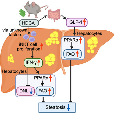
The researchers then identified two pathways through which HDCA activated PPARα. First, HDCA increased the number of invariant natural killer T (iNKT) cells, a type of immune cell, in the liver. These cells produced more interferon-γ (IFN-γ), a signaling molecule that activated PPARα in liver cells. PPARα then switched on the genes that help liver cells break down fatty acids.
As another pathway, HDCA also increased the levels of glucagon-like peptide-1 (GLP-1), a gut hormone that helps regulate metabolism. The researchers found that GLP-1 signaling was necessary for HDCA to reduce liver fat. When they tested mice that lacked the GLP-1 receptor, HDCA could no longer prevent fat from accumulating in the liver.
To further examine the role of GLP-1 and its relationship with iNKT cells and PPARα, the researchers injected liraglutide, a drug that activates the GLP-1 receptor and is used to treat type 2 diabetes, into normal mice and mice lacking iNKT cells, PPARα, or the GLP-1 receptor. Liraglutide reduced liver fat in both normal mice and mice lacking iNKT cells, but not in mice lacking PPARα. These findings indicated that GLP-1 reduces liver fat through PPARα and does not require iNKT cells.
Together, the researchers showed that HDCA may combat MASLD through two distinct pathways: an immune pathway involving iNKT cells and IFN-γ, and a metabolic pathway involving GLP-1. Both pathways ultimately activate PPARα, which helps the liver increase fatty-acid breakdown and reduce fat accumulation.
“Both HDCA and the GLP-1 receptor agonist liraglutide exhibited potent anti-steatotic effects in mice in a PPARα-dependent manner,” says Prof. Miki, highlighting PPARα as a potential target for developing strategies to combat MASLD.
Overall, these findings emphasize the potential of HDCA as an effective multi-target therapeutic strategy for the treatment of hepatic steatosis.