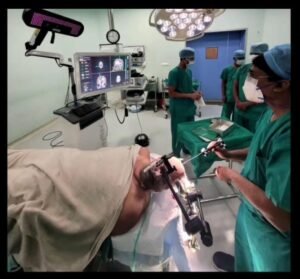
Mumbai- Solaris Hospital, Thane has carved a niche in Navigation Guided Brain and Spine Surgeries by introducing artificial intelligence (AI) driven Neuro- Navigation technology for the first time in Thane. Through this technology, Magnetic Resonance Imaging (MRI) or Computerized Tomography (CT) scans of patients can guide intraoperatively in real time for brain and spine surgeries towards effective clinical outcomes, safety, and medical management. “Neuro- Navigation technology is today playing a pivotal role in medical management with such newer advances.
Just like Navigation helps the pilot to find its path in the dark sky in real-time, Neuro Navigation guides the Neurosurgeon for the exact location of the tumor, its size, and extent, the limitation of critical structures, and also to minimize the size of craniotomy for Minimally Access Surgeries (MAS), informs Dr. Amit Aiwale, Neurosurgeon, Solaris Hospital, Thane. Solaris Hospital and Dr. Amit Aiwale is the first in Thane to introduce this technology in spine surgeries where CT guided Navigation is used for putting screws and implants in the spine. This improves the accuracy and precision of the surgeon.

Dr Amit Aiwale further explains that Neuro Navigation is the future wherein precision surgeries are in demand. Latest technologies like Neuro Navigation by surgeons should be adapted for better patient outcomes and satisfaction. “The benefits of computer Navigation Guided Brain and Spine Surgery include increased precision, enhanced safety, improved visualization, minimized radiation exposure, and enhanced training and education,” Dr Amit Aiwale explains about the USP of the technology. Increased precision implies that the technology allows for highly accurate targeting and placement of surgical instruments, minimizing the risk of damage to critical structures and improving surgical outcomes.
Enhanced safety implies that surgeons can navigate through complex anatomical structures with confidence, reducing the potential for complications and minimizing the invasiveness of the procedure. Improved visualization implies that the 3D virtual models and real-time imaging provide detailed and comprehensive visualization of the surgical site, aiding the surgeon’s decision-making process.
Minimized radiation exposure implies that computer navigation reduces the need for intraoperative imaging, reducing radiation exposure for both the patient and surgical team. The technology can be used for training purposes, allowing surgeons to simulate procedures and develop their skills in a controlled environment.
However, it is to be noted that computer navigation-guided surgery is a tool that complements the surgeon’s expertise and does not replace their judgment and skill. The surgeon remains in control of the procedure, using the navigation system as a valuable aid to improve surgical accuracy and patient outcomes.
How computer navigation guided brain and spine surgery works in a step-by-step manner is illustrated below. Preoperative Imaging: Before the surgery, the patient undergoes imaging scans such as MRI or CT scans. These scans provide detailed images of the patient’s brain or spine, allowing the surgeon to identify the target area and any abnormalities that need to be addressed.
Key Highlights
Image Registration: The imaging data is then loaded into the computer navigation system. The software matches the patient’s actual anatomy with the images from the scans, creating a three-dimensional (3D) virtual model of the patient’s brain or spine.
Reference Frame/ Point Placement: To establish a reference point, a frame with reflective markers or a specialized device is attached to the patient’s head or spine. These markers allow the computer system to track the patient’s movements during the surgery and accurately correlate them with the preoperative images.
Real-Time Tracking: Once the surgery begins, the computer navigation system tracks the position and movement of surgical instruments and the patient’s anatomy in real time. Specialized tools, such as infrared cameras or electromagnetic tracking systems, are used to monitor the position and orientation of instruments relative to the patient’s anatomy.
Surgical Guidance: The surgeon can visualize the patient’s anatomy, including the target area, surrounding structures, and critical landmarks, on a computer screen or a head-mounted display. This visualization helps the surgeon navigate precisely and avoid vital structures during the procedure.
Intraoperative Updates: During the surgery, the navigation system continuously updates the virtual model based on the tracked movements and instruments’ positions. This ensures that the surgeon has up-to-date information about the current state of the patient’s anatomy, even if there are minor changes or shifts during the procedure.
Verification and Validation: Throughout the surgery, the surgeon can validate the accuracy of their instrument’s position and orientation by referencing the virtual model and comparing it to the real-time imaging provided by the navigation system. This verification step ensures that the surgical plan is followed accurately.
Leave a Reply